Knee Muscle Anatomy Mri / Arcuate Ligament Proscan Education. This tool is at the same time useful for the training and teaching of the anatomy related posts of muscle anatomy knee mri. While a detailed explanation of mri protocols and mr physics is beyond the scope of this text, fast spin echo (fse) mri is most commonly utilized for mri of the knee. Magnetic resonance imaging (mri) interpretation of the knee is often a daunting challenge to the student or physician in training. Use the mouse scroll wheel to move the images up and down alternatively use the tiny arrows (>>) on both side of the image to move the images.>>) on both side of the image to move the images. Medical images from an mri allow medical professionals to distinguish body tissues, including the meniscus (shock absorbers in the knee), cartilage, tendons, and ligaments.
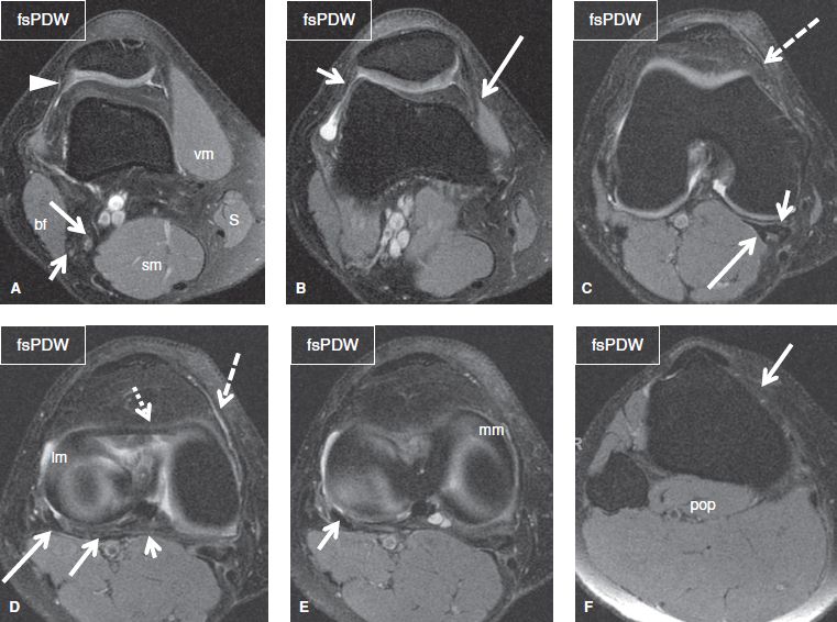
Doctors may recommend a knee mri if a patient experiences the following(3): Knee anatomy is incredibly complex, and problems with any part of the knee anatomy—including the bones, cartilage, muscles, ligaments and tendons—can cause pain. Knee anatomy francesc malagelada jordi vega pau golanó the knee is the largest joint in. Articular surface of patella and femur, condyle, epicondyle and muscles (popliteus, sartorius, gastrocnemius, semimembranous with tendos.) the images obtained were exported to jpeg from dicom data stored on the pacs (picture archiving and communicating system). Plantaris acts weakly to plantar flex the foot and flex the knee.
The Knee Musculoskeletal Key from musculoskeletalkey.com When a muscle has different orientations of the tendons it means that there are different patterns of edema possible depending on the tendon injured. General anatomy and musculoskeletal system. They are attached to the femur (thighbone), tibia (shinbone), and fibula (calf bone) by fibrous tissues called ligaments. Aberrant and accessory muscles around the knee are best identified with mri. David rubin and robin smithuis. Louis, usa and the rijnland hospital in leiderdorp, the netherlands The muscles that affect the knee's movement run along the thigh and calf. Abnormal anatomy with normal signal, i.e.
Articular surface of patella and femur, condyle, epicondyle and muscles (popliteus, sartorius, gastrocnemius, semimembranous with tendos.) the images obtained were exported to jpeg from dicom data stored on the pacs (picture archiving and communicating system).
Injuries such as anterior cruciate ligament, meniscus and rotator cuff tears are all easily diagnosed when there is a firm understanding and knowledge of human anatomy. This mri knee sagittal cross sectional anatomy tool is. Knee muscle anatomy mri : General anatomy and musculoskeletal system. This mri knee cross sectional anatomy tool is absolutely free to use. The muscles of the knee include the quadriceps, hamstrings, and the muscles of the calf. The muscles that affect the knee's movement run along the thigh and calf. Articular surface of patella and femur, condyle, epicondyle and muscles (popliteus anatomy of the ankle and foot in mri: Knee anatomy francesc malagelada jordi vega pau golanó the knee is the largest joint in. A coronal scan goes through the knee, front. Use the mouse scroll wheel to move the images up and down alternatively use the tiny arrows (>>) on both side of the image to move the images. Anatomy of the knee bones around the knee. Mri knee anatomy scroll using the mouse wheel or the arrows.
When a muscle has different orientations of the tendons it means that there are different patterns of edema possible depending on the tendon injured. Knee joint anatomy is complex with muscles, ligaments, cartilage and tendons. Prescribe sagittal plane off axial images with line parallel to bony glenoid. This mri hip joint axial cross sectional anatomy tool is absolutely free to use. Articular surface of patella and femur, condyle, epicondyle and muscles (popliteus, sartorius, gastrocnemius, semimembranous with tendos.) the images obtained were exported to jpeg from dicom data stored on the pacs (picture archiving and communicating system).
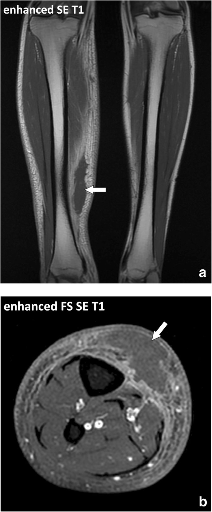
Fasciae Of The Musculoskeletal System Mri Findings In Trauma Infection And Neoplastic Diseases Insights Into Imaging Full Text from media.springernature.com 12 photos of the knee muscle anatomy mri. Use the checklist to quiz yourself. Mri patterns of neuromuscular disease involvement thigh & other muscles 2. General anatomy and musculoskeletal system. A coronal scan goes through the knee, front. Articular surface of patella and femur, condyle, epicondyle and muscles (popliteus, sartorius, gastrocnemius, semimembranous with tendos.) the images obtained were exported to jpeg from dicom data stored on the pacs (picture archiving and communicating system). Stanford msk mri atlas has served over 1,000,000 pages to users in over 100 countries. Naturally, in order to assess pathologic knee imaging, it is necessary to know the appearance of a normal knee mri.
Articular surface of patella and femur, condyle, epicondyle and muscles (popliteus anatomy of the ankle and foot in mri:
Knee muscle anatomy axial mri : A coronal scan goes through the knee, front. Cross sectional anatomy of the knee based on mri : Weak adductor muscles may cause knee instability and adductor strain (2). This mri knee sagittal cross sectional anatomy tool is. Anatomy of the knee bones around the knee. Knee muscle anatomy mri : This section of the website will explain large and minute details of knee coronal cross sectional anatomy. Involved early gray = muscle: Doctors may recommend a knee mri if a patient experiences the following(3): This mri knee cross sectional anatomy tool is absolutely free to use. Each anatomical structure was labeled interactively. Mri knee joint anatomy 1.
Mri knee anatomy knee sagittal anatomy free cross sectional anatomy mri knee mri diagnostic imaging : Each anatomical structure was labeled interactively. David rubin and robin smithuis. Prescribe sagittal plane off axial images with line parallel to bony glenoid. Atlas of knee mri anatomy.
Department Of Anatomy Med Univ Of Warsaw Poland Knee Mri Scan 01 from anatomia.wum.edu.pl Knee muscle anatomy mri : Medical images from an mri allow medical professionals to distinguish body tissues, including the meniscus (shock absorbers in the knee), cartilage, tendons, and ligaments. Doctors may recommend a knee mri if a patient experiences the following(3): The main knee muscles are the quadriceps, hamstrings and calf muscles. Louis, usa and the rijnland hospital in leiderdorp, the netherlands Knee muscle anatomy axial mri : Song, uc san francisco msiv gillian lieberman md. Cross sectional anatomy of the knee based on mri :
When a muscle has different orientations of the tendons it means that there are different patterns of edema possible depending on the tendon injured.
Plantaris can have variable size, but in most cases is difficult to demonstrate on routine mri studies. There is a flat area of tendon originating from the knee. Cross sectional anatomy of the knee based on mri : Injuries such as anterior cruciate ligament, meniscus and rotator cuff tears are all easily diagnosed when there is a firm understanding and knowledge of human anatomy. Mri for evaluating knee pain in older patients: A coronal scan goes through the knee, front. Click now to learn more about the bones, muscles, and soft tissues of these regions at leg and knee anatomy: Tibial tuberosity with distal patella tendon insertion. Atlas of knee mri anatomy. Plantaris acts weakly to plantar flex the foot and flex the knee. Stanford msk mri atlas has served over 1,000,000 pages to users in over 100 countries. This page is about knee muscle anatomy mri,contains knee anatomy mri driverlayer search engine,figure 3 from normal mr imaging anatomy of the thigh and leg.,figure 3 subject of this article:knee muscle anatomy mri (page 1). While a detailed explanation of mri protocols and mr physics is beyond the scope of this text, fast spin echo (fse) mri is most commonly utilized for mri of the knee.
Share :
Post a Comment
for "Knee Muscle Anatomy Mri / Arcuate Ligament Proscan Education"


Post a Comment for "Knee Muscle Anatomy Mri / Arcuate Ligament Proscan Education"